
By Dr. Cory Herman
TMJ patients who have a good understanding of their jaw anatomy tend to collaborate more effectively with their healthcare providers.
Having a basic knowledge of the anatomy of the jaw helps patients better communicate their symptoms, participate in treatment decisions, and actively manage their condition. The goal of this article is to help TMJ patients understand how their jaw muscles and joints work together.
Table of Contents
- Why Healthy Jaw Functioning Matters
- Key Parts of your Jaw’s Anatomy
- How the TMJ Works: Mechanics & Proper Function
- How Understanding Jaw Anatomy Helps Patients Manage TMJ
- Initial Jaw Anatomy Evaluation of TMJ Patients
- The Impact of Jaw Disc Displacement
- Education and Self-Management of Jaw Pain Contributes to Better Outcomes
- Understanding TMJ Dysfunction
- CONCLUSION: Holistic, Patient-Centered Approach to TMD Treatment
The temporomandibular joint (TMJ) is a complex hinge joint that connects your lower jaw (mandible) to your skull. Understanding what a healthy jaw joint anatomy looks like creates a visual goal when seeking proper functioning of the temporomandibular joints.
Why Healthy Jaw Functioning Matters
The TMJ’s connect your lower jawbone (mandible) to your skull. We each have 2 TMJ’s – one on the right and one on the left. These joints work together to allow for smooth movements like opening and closing your mouth, chewing, and speaking.
Proper jaw function is a result of the complex interaction between the jaw muscles, cartilage, and ligaments within the TMJ. This interaction allows for a wide range of motion, enabling you to comfortably perform everyday activities.
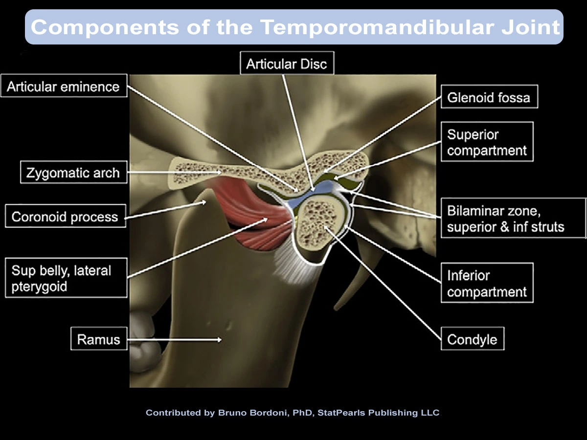
The key parts of your jaw’s anatomy include:
- Jaw Bones: The TMJ is formed by two bones: the mandible (your lower jawbone) and the temporal bone (a bone on the side of your skull).
- How Cartilage Supports Jaw Function: A fibrocartilaginous disc sits between these bones, acting like a cushion and allowing for smooth gliding movements.
- Ligaments Helping Jaw Function: Ligaments are strong, fibrous tissues that connect bones to each other and provide stability to the joint. Several ligaments support the TMJ.
- Muscles that Impact Jaw Movement: A network of muscles is responsible for controlling jaw movements. The primary muscles are the masseter, temporalis and pterygoid muscles.
How the TMJ Works: Mechanics & Proper Function
Key muscles operating your jaw include:
- Masseter: A powerful muscle that closes the jaw for chewing.
- Temporalis: A muscle that helps close the jaw and move it backward.
- Lateral pterygoid: This muscle helps open the jaw and move it forward.
- Medial pterygoid: This muscle helps close the jaw and move it sideways.
Trigeminal nerve anatomy:
The trigeminal nerve (cranial nerve V) controls the muscles of jaw movement and transmit sensations like pain to the brain. There are 3 branches of the trigeminal nerve, each of which need to function for movement and sensation of the jaw to occur properly.
When patients understand the rationale behind their pain treatment plan, they are more likely to adhere to it. If you have further questions about your jaw bones, cartilage, ligaments, and associated muscles, please contact us.
Now that we’ve explored the basic anatomy, let’s examine how these parts work together.
How your TMJ hinge works
When working at their best, jaw components should move smoothly and in coordinated fashion. This lets you enjoy effortless opening and closing with proper alignment of teeth, minimal strain on muscles, and no discomfort in the temporomandibular joints (TMJs). Essentially, a normal functioning jaw hinge gives you a balanced and efficient chewing motion with a good bite alignment.
Imagine your TMJ as a well-oiled hinge. When you open your mouth, the condyle (a rounded part of the mandible) rotates and glides forward within the socket of the temporal bone, facilitated by the disc and the muscles working in harmony. Closing your mouth involves the reverse action.
Maintaining a relaxed jaw posture is crucial for TMJ health. When your jaw is relaxed, the muscles are at rest, and the joint is not under constant pressure. Many people unknowingly engage in teeth clenching or grinding, putting excessive strain on the TMJ hinge and surrounding muscles.
By teaching the mechanics of your jaw movement, we make it easier for you to follow your provider’s instructions. The Minnesota Head & Neck Pain Clinic strives to ensure proper healing and optimal outcomes of your treatment plan. Below are specific ways that partnering with your provider helps.
Connective tissue, blood supply, and fascia:
The human body is so intricately made that issues in other areas of the body can contribute to jaw problems. Fascia in your neck and jaw region may form restrictions or adhesions that can affect jaw movement and pain. It is a type of connective tissue that surrounds blood vessels, organs, and other structures in your fascia. Movement limitations can occur in opening, closure, protrusion, and lateral excursion of the mandible.
The primary blood supply to your jaw includes the muscles used for jaw movement and comes from the maxillary artery. This is a branch of the external carotid artery; specifically, the inferior alveolar artery, which is a terminal branch of the maxillary artery. It is responsible for most of the blood supply to the mandible and surrounding tissues, including the teeth and nerves in the lower jaw.
Kushagra Maini explains in the January 30, 2023 Temporomandibular Syndrome article, “Three major ligaments stabilize the TMJ: temporomandibular, stylomandibular, and sphenomandibular ligaments.”
How Understanding Jaw Anatomy Helps Patients Manage TMJ
Understanding your TMJ anatomy helps with prevention by:
- Identifying pain source: Recognizing which muscles and ligaments are involved in one’s pain can help patients understand and articulate the source of their discomfort.
- Identifying risk behaviors: Knowing how the joint works helps you identify habits that put stress on the TMJ. Like, chewing gum excessively, teeth clenching or grinding, nail biting, poor posture, or opening your mouth too wide for too long. For example, you can ask your dentist for shorter appointments or to have breaks to avoid extended mouth opening.
- Self-management techniques: Knowing the importance of maintaining a relaxed jaw and continuous, proper and movement can inspire patients to persist in giving the detailed attention needed to correctly practice targeted jaw relaxation exercises. Additionally, maintaining good posture helps relieve jaw and neck pain. A forward head posture, rounded shoulders, or other postural imbalances can affect jaw alignment and contribute to muscle tension.
- Overall treatment compliance: A deeper understanding of TMJ mechanics can improve adherence to treatment plans. This may include simple things like the importance of engaging in clinical visits, attention to instructions for wearing a mouth guard, or faithfully performing physical therapyphysical therapy exercises.
- Principles of maxillofacial treatment: When it comes to diagnosing maxillofacial conditions of the jaw, TMJ, or oral medicine conditions, CBCT imaging can also play a crucial role. It not only helps providers establish a treatment plan, but it also lets patients visualize where issues may lie and where improvements are needed.
By being aware of the jaw joint’s mechanics, individuals can consciously engage in a mindfulness practice of jaw movements to avoid overexertion. Let’s quickly summarize this section before talking about what to expect during an examination of your jaw’s anatomy.
Key points about the TMJ joint:
- Location: In front of the ear.
- Function: Connects the lower jaw (mandible) to the skull (temporal bone).
- Number of joints: Two TMJ joints, one on each side of the head.
“Several ligaments manage the TMJ forces and send multiple proprioceptive afferents. The proprioception of the joint is provided by various components, such as the capsule, masticatory muscles, skin receptors and receptors within the periodontal ligaments. The tension perceived by the articular ligaments plays an important role in the function of TMJ.
“Its position and structure make it an intersection of information and influences that expand throughout the body, and vice-versa; the mechanical information of other body districts can reflect in the TMJ.” – NIH: Anatomy, Head and Neck, Temporomandibular Joint
Initial Jaw Anatomy Evaluation of TMJ Patients
As part of a complete assessment of jaw complaints, your Orofacial Pain specialist will perform an examination of your jaw structures and your jaw anatomy. This examination should be completed prior to any treatment of TMJ concerns as this will help to ensure that a proper diagnosis is made.
Things Orofacial Pain Specialists look for when assessing someone’s jaw anatomy:
- Jaw movement including measurement of jaw range of motion.
- Swelling of the jaw or TMJ’s and tenderness to touch of the jaw joints.
- Muscle tenderness or trigger points (i.e. muscle knots).
- An evaluation of the teeth and bite to look for evidence of teeth clenching or grinding.
- Evaluation of the proper function of the cranial nerves of the jaw and face.
- A screening dental examination to rule out any obvious dental or gum issues.
Understanding how the TMJ should function is crucial; let’s now explore what happens when that function is disrupted.
The Impact of Jaw Disc Displacement
Jaw disc displacement is a very common issue in TMJ disorders and directly relates to clicking, popping, and limited range of motion. It impacts the joint’s functional and can cause pain. The disc within the TMJ can become displaced, disrupting the smooth movement of the joint and causing pain.
“Disc displacement with reduction” refers to a condition where the TMJ disc temporarily moves out of place but then returns to its normal position during jaw movement. This often causes a clicking or popping sound, while “disc displacement without reduction” means the disc remains displaced, preventing full jaw opening and causing limited mouth movement without a noticeable clicking sound.
Disc displacement with reduction is often considered a milder condition compared to without reduction, which can cause a higher levels of jaw pain and functional limitations. Internal derangements is another name for a disc displacement.
Let’s try simplifying this. With reduction, the disc “clicks back into place” while opening the mouth, whereas without reduction, the disc stays displaced, limiting jaw opening.
You may be wondering, why does this matter to you?
An altered jaw anatomy such as a disc displacement can occur as a result of repetitive strain (i.e. teeth clenching or poor posture) or trauma to the temporomandibular joint. This could be due to a sports injury to the head and neck region, which can lead to conditions like TMDs. If the cartilage and surrounding soft tissues within your jaw joint becomes irritated or damaged due to excessive strain, or injury, pain may result. The pain may be accompanied with jawlock, and limited jaw movement; this can also affect the muscles controlling the jaw, causing further discomfort and tissue damage.
Understanding potential tissue damage from jaw dysfunction can help guide you through pain management.
Potential tissue damage from jaw anatomy issues:
- Muscle strain and spasms: Overworked jaw muscles due to teeth clenching or grinding can trigger jaw strain and muscle spasms, leading to pain and restricted jaw movement.
- Ligament damage: The ligaments supporting the TMJ can become stretched or torn due to trauma or excessive jaw movement.
- Cartilage erosion: Continuous stress on the joint can wear down the protective cartilage lining, causing joint pain and inflammation.
- Nerve irritation: The trigeminal nerve, which supplies sensation to the face, can be irritated by a dysfunctional TMJ, leading to myofacial pain and headaches.
- TMD: Temporomandibular disorders encompass a group of conditions that cause pain and dysfunction. Sometimes, the leading cause is excessive strain triggered by jaw overuse that impacts the muscle group that controls chewing, swallowing, and speech.
TMD may be muscle-related, joint-related, disc-related, or even from an arthritic complication in the jaw area. Not all TMJ pain is the same. Various causes require different approaches. Potential causes, triggers, nerve pathways and pain sensitization are carefully assessed. Remember, open dialog about what you feel helps our diagnosis and helps with managing expectations.
Education and Self-Management of Jaw Pain Contributes to Better Outcomes
The Minnesota Head & Neck Pain Clinic strongly believes in patient involvement. We want to empower you to take an active role in your treatment.
On the clinical side, it’s our belief that education and self-management strategy for patients with TMD should rely on an evidence-based approach to jaw pain treatment.
“Education based on a neuroscience approach, highlighting the differences between pain and nociception, emphasizing that the injured tissue is neither necessary nor sufficient to perceive pain, and providing a list of contributing factors that could perpetuate the symptomatology such as negative thoughts, emotions, anxiety or scant physical activity, has shown good results in pain relief, reducing psychosocial factors and disability in patients with chronic musculoskeletal pain.” – Temporomandibular disorders: improving outcomes using a multidisciplinary approach
The above article references James Fricton DDS, MS stating that “the most frequent cause of therapeutic failure is a bad diagnosis.” It is a mistake to try to treat TMD as a single disease.
Understanding TMJ Dysfunction
Within the jaw anatomy, the TMJ is a complex joint.
TMJ pain isn’t always localized to the jaw joint itself. Chronic pain can alter your nervous system (sensitization); that is why pain can sometimes be disproportionate to tissue damage. When patient’s collaborate with providers who take a team approach, your Orofacial Pain Specialist can help you understand referred pain and pain exacerbations.
Referred pain
The brain can sometimes misinterpret pain signals, leading to pain felt in areas other than the source. Jaw joint and jaw muscle pain can cause pain referral and be felt in distant areas, such as headaches, earaches, neck pain, shoulder pain, and tooth pain. This can make it challenging to pinpoint the problem’s root cause without a specialist’s expertise.
Pain exacerbations
If your jaw anatomy is negatively impacted, pain may not be constant and can fluctuate in intensity. “Pain exacerbations” are periods when pain intensifies. An Orofacial Pain Specialist has the training and expertise to identify the true source of pain (including referred pain) and help you manage chronic pain and its associated flare-ups.
You should be actively involved in your treatment decisions and have access to the information and resources you need to make informed choices. We encourage questions and actively listen to your concerns in order to best address individual needs.
CONCLUSION: Holistic, Patient-Centered Approach to TMD Treatment
We’ve covered the physical structure of the human jaw; however, we know that reducing TMJ pain is often more complex. A holistic approach to solving jaw issues means considering the patient’s individual needs and preferences and addressing any underlying psychological or social factors that may be contributing to their condition. Listen to our TMD podcast to learn more.
Schedule Your Jaw Joint Examination
About the Author
Cory Herman DDS, Doctor of Dental Surgery, treats adults and children with sleep issues, TMJ, and Orofacial pain disorders within the Minnesota Head and Neck Pain Clinic. He is known for his non-surgical jaw pain management with an emphasis on collaborative and integrative care.
Medical research conducted by Hill Web Marketing.